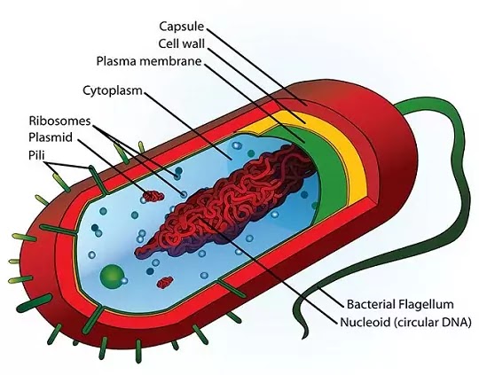
Bacteria are unicellular organisms. These are very small in size and thus little can be know about them by conventional light microscope. Their fine structure has been revealed with the help of electron microscope. All bacteria show typical prokaryotic structure. Like other plant cells, in bacteria protoplast is covered by cell wall.
1. The outermost covering of slime
2. The middle cell wall
3. Innermost cytoplasimic membrane
1. Slime / Capsule layer - It is a gelatinous layer present on the outer surface of the cell wall, It is composed of polysaccharides (example - dextran, dextrin, lavan) or polypeptide chains of amino acids. When its constituents are only polysaccharides, which form a viscous layer, it is said to be slime layer but when nitrogenous substances (i.e., amino acids) are also present along with polysaccharides, then it is called capsule. Various strains of the same species may have capsules of several different strain specific polysaccharides. The slime layer/capsule protects the cell from desiccation and antibodies. The capsulated bacteria also unaffected by phagocytosis. They are usually atrichous.
2. Cell wall - Each bacterial cell remains surrounded by a thick and rigid cell- wall. It is usually present outside the cell- membrane. Its thickness varies from 50Å to 100 Å.
The chief functions of cell-wall are-
(i) to provide definite shape to the cell,
(ii) to protect protoplasm from adverse conditions,
(iii) to control the entry and exit of molecules of various substances.
(iv) to control the entry or exit of large sized molecules of proteins, nucleic acid and viruses etc.
Chemically composed of Bacterial cell wall is
(i) N - acetylglucosamine (AGA)
(ii) N-acetye muramic acid (AMA) also called NAM. NAM provides rigidity. NAM and NAG are linked alternately by B, 1, 4-glycosidic bonds.
(iii) Amino acids - The peptidogly can chains are laterally linked by short chain of four amino acids which are attached to NAM. The four amino acids of this tetrapeptide are (a) D-alanine (b) L-alanine (c) D-glutamic acid (d) L-lysine in Gram (+) and diaminopimellic acid in Gram (-). Mucopeptide or peptidoglycan or murien Peptidoglycon.
The cell wall of gram positive bacteria is characterized by the presence of teichoic acid (acid polymers having glucose, phosphate and alcohol). Teichoic acid acts as receptor site and attract chemicals that provide protection from thermal and pH changes. Cellulose is generally absent from the cell wall of bacteria but Thimann reports its presence in Azotobacter and Zymosarcinu. In some bacteria example- Mycobacterium cell well contains mycolic acid.
3. Cytoplasmic membrane - It is just below the cell wall and semi permeable cytoplasmic membrane is present. Which is about 75Å thick. Chemically it is composed of a double layer of phospholipid molecules. Phospholipids are of two types hydrophobic and hydrophilic. The hydrophilic phospholipid molecules are present towards the outerside and the hydrophobic molecules towards the inner side. Proteins are found embedded in the lipid bilayer. Prokaryotic membrane is characterized by the absence of sterols which perhaps account for the enormous resistance of these organisms to antibiotics. The chemical nature of cytoplasmic membrane in Gram-positive and Gram-negative bacteria is basically the same.
Cytoplasmic membrane is the bounding layer of the cytoplasm and it is the centre of multifarious activities of the cell. Enzymes of various metabolic pathways, such as synthesis of lipopolysaccharides, phospholipids, teichoic acid, etc., are located in the cytoplasmic membrane. Space between cell wall and cytoplasmic membrane is called periplasmic space.
4. Cytoplasm - The bacterial cytoplasm is a complex mixture of carbohydrates, proteins, lipids, minerals, nucleic acids and water. It stores organic material in the form of glycogen, volutin and poly- ẞ-hydroxybutyrate. Some photosynthetic and non-photosynthetic bacteria also accumulate sulphur and iron in their cytoplasm. The bacterial cell is devoid of mitochondria, endoplasmic reticulum, centrosome and Golgi bodies. Chloroplast is absent in bacteria. Photosynthetic bacteria chromatophores in their cytoplasm.
5. Nuclear Apparatus - Definite, particulate nucleus is absent in bacteria. The bacteria possess primitive type of nuclear apparatus which does not have nuclear envelope, nucleolus, chromonemata and nuclear sap. It divides amitotically. Such nucleus is termed as nucleoid, chromatin body or genophore. Under electron microscope the nuclear region appears as an electron translucent structure and contain very fine fibrils. These fibrils are molecular strands of single or double stranded DNA.
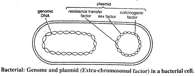
6. Plasmids - Extrachromosomal hereditary of Bacterial cells contain and determinants which are either independent of bacterial chromosomes or are integrated with them. Lederberg (1952) called the term plasmids for such extrachromosomal hereditary determinants. They are rings of DNA of various sizes. Each plasmid carries non-essential genes and has no role in viability and growth of bacteria. Hence they are also called to be dispensable autonomous elements. One good example of plasmids is the F-factor which determines the maleness in bacteria. It is an autonomous element, separate from the bacterial chromosome. It is transmitted from cell to cell by contact or by external agencies. A number of bacterial properties are now known as plasmid carried genes. Plasmids have been classified on the basis of the host properties.
The following nine types of plasmids are known
(i) F-factor (for fertility) plasmids,
(ii) R-factor, (for resistance) plasmids,
(iii) col-factor (for colicinogeny) plasmids of the Gram-negative bacteria,
(iv) plasmids conferring pathogenecity to mammals,
(v) degradative plasmids of Pseudonionas,
(vi) mercury-resistance plasmids,
(vii) tumor inducing plasmids of Agrobacterium tumifaciens,
(viii) penicillase plasmids of Staphylococcus aureus,
(ix) cryptic plasmids.
Circular double stranded DNA molecules found in plasmid. Their molecular weight ranges between 5 x 10⁷ and 7 x 10⁷ (some cryptic plasmids are even smaller). Each plasmid may contain as many as 100 genes. The general features of plasmid replication are similar to that of chromosome replication, but plasmid seems to operate under its own genetically determined system of replication control.
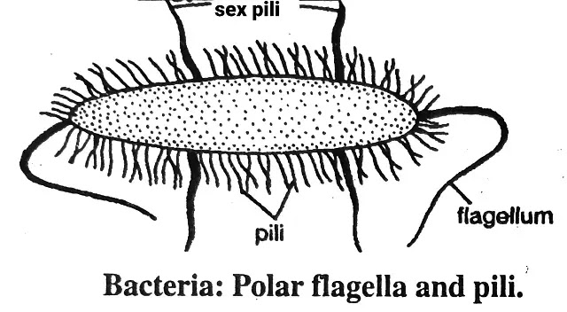
7. Motility or Flagella - Bacteria are motile as well as non-motile. Movement in bacteria takes place by appendages called flagella. They are extremely long and thin. The flagella are presenting in motile and absent in non-motile forms of bacteria. When the flagellum rotates clockwise, the bacterium moves forward. When the rotation is anticlockwise, the bacterium tumbles about chaotically.
Bacteria are divided into the following forms -
(I) Atrichous - When the flagellum is absent. These bacteria are non-motile, example- Pasteurella.
(II) Trichous - Such bacteria possess flagella and are motile. On the basis of number, position and arrangement, these are further divided into following categories give below -
(a) Polar Flagella - Here flagella are restricted to the ends of the bacteria. Usually Gram-negative bacilli and spiral bacteria have such flagellation. Polar flagella is further divided into following categories -
- Monotrichous - When there is only one flagellum on one end of the bacterium, example- Vibrio. Pseudomonas.
- Lophotrichous - When there is group of two or more flagella at one pole of the cell, they are called as lophotrichous. Example- Spirillum serpens.
- Amphitrichous - When there is a atleast one flagella on both the ends of the bacterium, they are called as Amphitrichous. example - Nitrosomonas.
(B) Non-polar Flagella - Here flagella are not restricted to the ends of bacteria.
Peritrichous - When flagella is found on the whole body of bacterium, These type flagella are called as Peritrichous e.g - Proteus vulgaris.
Structure of Bacterial flagellum
Structure of bacterial flagella are quite unlike those of other organisms. The flagella and cilia of higher protists and other organisms have an identical 9 +2 fibrillar structure. The molecular architecture of bacterial flagellum, derived on the basis of chemical analysis, reveals that it is composed of several chains of flagellin (a protein) molecules forming a more or less cylindrical filament. The diameter of a flagellin molecule is about 40Å and that of a flagellum is about 120-150 Å. The orientation of flagellin molecules may vary in different species. The length of flagella (4-5 μm long) is usually more than that of bacterial cell. The swimming ability of bacteria is due to the presence of flagella. If the cell is mechanically deflagellated, it becomes immotile. The locomotion is brought about by clockwise or anticlockwise spins of flagellum around its own 'axis'. In a liquid medium the speed of movement is more than 20 tm per second.
8. Pili or fimbriae - These are straight hair like structure which are much shorter and thinner than the flagella. They cover the body of the cell and there may be 100-400 of them on one cell. Pill are 0.3 - 10 μ long and 0.01 μ wide. It is supposed that these are not related to the locomotion of the cell and that they serve to attach the bacterial cells to the surface of some substrates. These consist of special type protein called 'Pilin'. Just like in the case if flagella, it is not necessary that all bacterial cells have pili.
8. Pili or fimbriae - These are straight hair like structure which are much shorter and thinner than the flagella. They cover the body of the cell and there may be 100-400 of them on one cell. Pill are 0.3 - 10 μ long and 0.01 μ wide. It is supposed that these are not related to the locomotion of the cell and that they serve to attach the bacterial cells to the surface of some substrates. These consist of special type protein called 'Pilin'. Just like in the case if flagella, it is not necessary that all bacterial cells have pili.
9. Gram's Stain - The procedure involves staining the cells first with crystal violet and then with iodine solution. The group of bacteria which retain the stain even after decolonization with alcohol is called Gram-positive, whereas those which lose stain after the treatment with alcohol are called Gram-negative. This differential reaction of two types of bacteria to crystal violet-iodine stain is due to different amount of lipids in their cell wall. The Gram-negative bacteria have relatively high lipid content. Alcohol dissolves the lipid which allows the leakage of stain. The Gram-positive bacteria, on the other hand, have less lipids and hence less susceptible to alcohol action. Gram's stain has long been used to separate bacteria into two groups, the gram-negative and positive types.


No comments:
Post a Comment